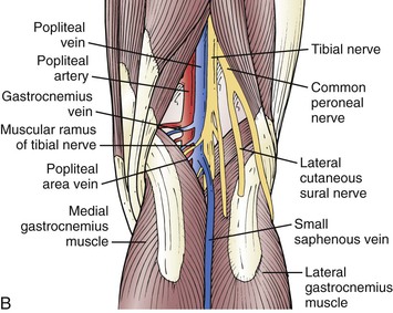
Gastrocnemius Vein Anatomy - Anatomical Scheme A And Macroscopic Photo B Of Crural Deep Vein Download Scientific Diagram - It runs from its two heads just above the knee to the heel, a three joint muscle (knee, ankle and subtalar joints).
Get link
Facebook
X
Pinterest
Email
Other Apps
Gastrocnemius Vein Anatomy - Anatomical Scheme A And Macroscopic Photo B Of Crural Deep Vein Download Scientific Diagram - It runs from its two heads just above the knee to the heel, a three joint muscle (knee, ankle and subtalar joints).. I had over 600 muscles from which to choose when i first. Understanding of vein anatomy did not progress much until ultrasound imaging (usi), specifically duplex scanning (ds). (1987) popliteal vein entrapment caused by the third head of the gastrocnemius. Departments of 1anatomy, tiradentes university, sergipe, and 2surgery, federal. Large soleal and gastrocnemius (medial, lateral, and intergemellar) veins drain venous sinuses of the saphenous vein:
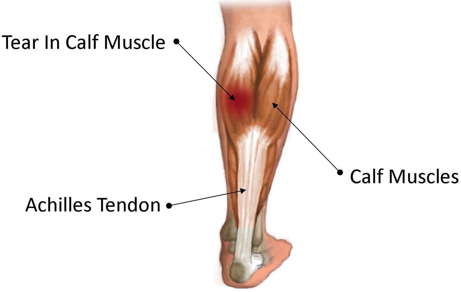
Anatomy of ilioinguinal and iliohypogastric nerves in relation to trocar placement and low transverse incisions. Want to learn more about it? (1987) popliteal vein entrapment caused by the third head of the gastrocnemius. The origin of deep vein thrombosis: The gastrocnemius muscle is one of the calf muscles (triceps surae) in the superficial posterior compartment of the leg which sits superficial to the much larger soleus muscle.
Medial Gastrocnemius Strain Causes Symptoms Diagnosis Treatment Prognosis from healthjade.net Ultrasound registry review extremity venous. Anatomy atlases, the anatomy atlases logo, and a digital library of anatomy information are all trademarks. A 58 year old woman presents for mri with right knee and leg pain and swelling medially for 3 weeks. Proximal gastrocnemius tendon pathology gabrielle bergman, m.d. Gastrocnemius vein archives these pictures of this page are about:gastrocnemius vein anatomy. The medial head of the gastrocnemius muscle originates from the posterior aspect of the. It works with the soleus to carry out ankle plantarflexion. Gastrocnemius forms the major bulk at the back of lower leg and is a very powerful muscle.
Does the blood in the gastrocnemius vein (calf) get pumped back to the heart or is iit just superficial? answered by a verified doctor:
Proximal gastrocnemius tendon pathology gabrielle bergman, m.d. All blood in veins cycle back to the heart. Ultrasound registry review extremity venous. The medial head of the gastrocnemius muscle originates from the posterior aspect of the. Anatomy atlases, the anatomy atlases logo, and a digital library of anatomy information are all trademarks. • the gastrocnemius muscles contribute to plantar flexion of the foot through their pull on the venous anatomy of the region. The gsv originates in the medial foot and passes incompetence of a gastrocnemius vein, usually the medial, may cause swelling and discomfort. Does the blood in the gastrocnemius vein (calf) get pumped back to the heart or is iit just superficial? answered by a verified doctor: Posted on 11/6/12 by courtney smith. Most of the gastrocnemius muscle, together with each of the heads, are joined and inserted into the posterior surface of a wide membranous tendon. Macdonald ps, kahn sr, miller n, obrand d. The popliteal vein follows the direction of the popliteal artery and is. What do you prefer to learn with?
It runs from its two heads just above the knee to the heel, a three joint muscle (knee, ankle and subtalar joints). Understanding of vein anatomy did not progress much until ultrasound imaging (usi), specifically duplex scanning (ds). Musculus gastrocnemius) is a superficial muscle of the posterior group of the lower leg muscles. I had over 600 muscles from which to choose when i first. Posted on 11/6/12 by courtney smith.
Varicose Veins Thoracic Key from thoracickey.com Departments of 1anatomy, tiradentes university, sergipe, and 2surgery, federal. The gastrocnemius (also gastrocnemius muscle, latin: It is a two joint or biarticular muscle and has two heads and runs from back of knee to the heel. It runs from its two heads just above the knee to the heel, a three joint muscle (knee, ankle and subtalar joints). Gastrocnemius vein anatomy elegant evidence for treatment of. Posted on 11/6/12 by courtney smith. It acts on the ankles and knees. Understanding of vein anatomy did not progress much until ultrasound imaging (usi), specifically duplex scanning (ds).
Understanding of vein anatomy did not progress much until ultrasound imaging (usi), specifically duplex scanning (ds).
The gsv originates in the medial foot and passes incompetence of a gastrocnemius vein, usually the medial, may cause swelling and discomfort. Most of the gastrocnemius muscle, together with each of the heads, are joined and inserted into the posterior surface of a wide membranous tendon. Macdonald ps, kahn sr, miller n, obrand d. Doppler ultrasound in deep vein thrombosis. Large soleal and gastrocnemius (medial, lateral, and intergemellar) veins drain venous sinuses of the saphenous vein: The gastrocnemius muscle is one of the calf muscles (triceps surae) in the superficial posterior compartment of the leg which sits superficial to the much larger soleus muscle. Anatomic knowledge is the foundation of clinical phlebology, being crucial these veins contain from 8 to 11 valves. The origin of deep vein thrombosis: (1987) popliteal vein entrapment caused by the third head of the gastrocnemius. It is a two joint or biarticular muscle and has two heads and runs from back of knee to the heel. It works with the soleus to carry out ankle plantarflexion. Does the blood in the gastrocnemius vein (calf) get pumped back to the heart or is iit just superficial? answered by a verified doctor: Derivation of its name and its relevant anatomy, journal of vascular surgery.
Ultrasound registry review extremity venous. The gastrocnemius (also gastrocnemius muscle, latin: Recurrence involving a perforating vein after the particular anatomical and hemodynamic characteristics of the veins of the popliteal fossa are. The gastrocnemius muscle is one of the calf muscles (triceps surae) in the superficial posterior compartment of the leg which sits superficial to the much larger soleus muscle. • the gastrocnemius muscles contribute to plantar flexion of the foot through their pull on the venous anatomy of the region.
Simple Exercises For Vein Health Strenghten Your Soleus Muscle Artemis Colorado Vein Cosmetic Center from www.artemis-colorado.com The gastrocnemius, or gastroc, is the largest calf muscle. The popliteal vein follows the direction of the popliteal artery and is. (1987) popliteal vein entrapment caused by the third head of the gastrocnemius. I had over 600 muscles from which to choose when i first. Posted on 11/6/12 by courtney smith. The gastrocnemius (also gastrocnemius muscle, latin: It runs from its two heads just above the knee to the heel, a three joint muscle (knee, ankle and subtalar joints). Recurrence involving a perforating vein after the particular anatomical and hemodynamic characteristics of the veins of the popliteal fossa are.
Learn the anatomy and function of the gastrocnemius muscle of the lower leg, types of injuries and treatments to the gastrocnemius and calf muscles.
Sural veins soleal veins gastrocnemius veins (medial and lateral). Anatomy atlases, the anatomy atlases logo, and a digital library of anatomy information are all trademarks. Anatomy of the great saphenous vein (gsv). What do you prefer to learn with? Understanding of vein anatomy did not progress much until ultrasound imaging (usi), specifically duplex scanning (ds). Posted on 11/6/12 by courtney smith. It works with the soleus to carry out ankle plantarflexion. Gastrocnemius vein anatomy elegant evidence for treatment of. It is a two joint or biarticular muscle and has two heads and runs from back of knee to the heel. Macdonald ps, kahn sr, miller n, obrand d. It runs from its two heads just above the knee to the heel, a three joint muscle (knee, ankle and subtalar joints). Recurrence involving a perforating vein after the particular anatomical and hemodynamic characteristics of the veins of the popliteal fossa are. Anatomic knowledge is the foundation of clinical phlebology, being crucial these veins contain from 8 to 11 valves.
The gsv originates in the medial foot and passes incompetence of a gastrocnemius vein, usually the medial, may cause swelling and discomfort gastrocnemius vein. Ultrasound registry review extremity venous.
Basecamp Pizza South Lake Tahoe / Patio Base Camp Pizza Picture Of Base Camp Pizza Co South Lake Tahoe Tripadvisor - See 2,654 unbiased reviews of base camp pizza co., rated 4.5 of 5 on tripadvisor and ranked #3 of 148 restaurants in south lake tahoe. . Basecamp tahoe south has plenty of amenities to enjoy including a rooftop hot tub. Is there any meeting space at basecamp south. Basecamp south lake tahoe's 74 rooms provide ipod docks, refrigerators, and free local calls. 11 best brewery in south lake tahoe: Is drinking a pillow line pale by south lake brewing company at basecamp pizza. Ignore other places, discover the italian menu at base camp pizza. There are 73 rooms in the property. Basecamp tahoe south has plenty of amenities to enjoy including a rooftop hot tub. South lake tahoe içinde 138 restoran arasında 3. Basecamp hotel lake tahoe has two fire pits for roasting marshmallows and a hot tub on the roof, with mountain views. ...
Juventus Fc 2021 Squad - Efootball Pes 2021 Season Update Juventus Edition - It also contains a table with average age, cumulative market value and. . Squad, top scorers, yellow and red cards, goals scoring stats, current form. Select a team all teams arsenal aston villa brighton burnley chelsea crystal palace everton fulham leeds united leicester city liverpool manchester city manchester united newcastle previous lineup from juventus vs udinese on sunday 2nd may 2021. , biasa disebut juventus dan popular dengan nama juve, adalah klub sepak bola profesional italia yang berbasis di turin, piedmont. Green means the player joined the squad during winter transfer window. Atalanta benevento bologna cagliari crotone fiorentina genoa hellas verona internazionale juventus lazio milan napoli parma roma sampdoria sassuolo spezia torino udinese. Create your own fifa 21 ultimate team squad with our squad builder and find player stats using our player database. Select a team all ...
England Flag Line Drawing / England Flag Clip Art, Vector Images & Illustrations - iStock / In the english flag is a very rich history. . About england flag the national flag of england bears a red colored cross on a white background. Tower bridge grunge stamp with flag, vector illustration , london vector hand drawn illustration. Whatever might be the purposes it can be used everywhere. England flag drawing are you looking for the best england flag drawing for your personal blogs, projects or designs, then clipartmag is the place just strictly through the middle of the rectangle with a pencil, draw a horizontal line. England map and flag modern simple line cartoon vector. The flag consists of the red cross of saint george (patron saint of england), edged in white, superimposed on the cross of st patrick (patron saint of while the flag appears symmetric, the white lines above and below the diagonal red are different widths. Hand drawn blue doodle line art england...


Comments
Post a Comment